HAEMOPHILUS INFLUENZAE & OTHER HAEMOPHILUS SPECIES
Essentials of Diagnosis
- Haemophilus influenzae is generally acquired via the aerosol route or by direct contact with respiratory secretions.
- The most common associated syndromes include otitis media, sinusitis, conjunctivitis, bronchitis, pneumonia, and, to a lesser extent, meningitis, epiglottitis, arthritis, and cellulitis.
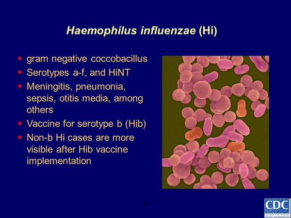
- Gram stain shows pleomorphic gram-negative coccobacilli.
- In cases of meningitis, epiglottitis, arthritis, and cellulitis, organisms are typically recovered from blood, and type-b polysaccharide capsular material may be detected in the urine.
- Organisms and type-b polysaccharide capsule may also be present in other appropriate sterile body fluids, such as cerebrospinal fluid (CSF) in meningitis and joint fluid in arthritis.
General Considerations
Epidemiology
Before 1990, strains of Haemophilus influenzae type b were found in the upper respiratory tract of 3-5% of children and a small percentage of adults. Colonization rates with type-b strains are even lower now, reflecting routine immunization of infants against H influenzae type b. Non-type-b encapsulated H influenzae are present in the nasopharynx of < 2% of individuals, whereas nonencapsulated (nontypable [see below]) strains colonize the respiratory tract of 40-80% of children and adults.

Historically, H influenzae type b was the leading cause of bacterial meningitis and epiglottitis in children < 5 years old and a major cause of septic arthritis, pneumonia, pericarditis, and facial cellulitis in this same age group. In the United States, ~ 1 in 200 children experienced invasive (bacteremic) disease with this organism before the age of 5 years, with a peak incidence of disease at 6-7 months of age. Invasive disease was more frequent in boys, children of African descent, Alaskan Eskimos, Apache and Navajo Indians, child care center attendees, and children living in overcrowded conditions. Other factors predisposing to invasive disease included sickle cell disease, asplenia, human immunodeficiency virus (HIV) infection, certain immunodeficiency syndromes, and malignancies. The introduction of efficacious vaccines and their routine use in infants, beginning in 1991, resulted in a marked decrease in the incidence of H influenzae type-b infections, which now are quite rare. The Immunization Practices Advisory Committee of the Centers for Disease Control and Prevention and the Committee on Infectious Diseases of the American Academy of Pediatrics currently recommend administration of a licensed conjugate vaccine to all children starting at 2 months of age. Invasive disease in this country now occurs primarily in undervaccinated children.
Nonencapsulated strains of H influenzae are a common cause of localized respiratory tract disease in both children and adults. In children, these organisms are the most common cause of purulent conjunctivitis, the second most common cause of otitis media (after Streptococcus pneumoniae), and a frequent cause of sinusitis. Among children in developing countries, they are a frequent cause of pneumonia and an important source of mortality. In adults they are especially common as a cause of community-acquired pneumonia and exacerbations of underlying lung disease and also account for ~ 30% of cases of otitis media and sinusitis. Beyond producing localized disease, nontypable H influenzae is an occasional cause of serious systemic disease, such as sepsis, meningitis, and pyogenic arthritis, particularly in neonates and individuals with compromised immunity.
In the mid-1980s, H influenzae biogroup aegyptius was recognized as the etiology of Brazilian purpuric fever (BPF), a septicemic illness occurring in young children and associated with a case fatality rate of ~ 60%. In most cases, disease is preceded by purulent conjunctivitis. Both epidemics and sporadic cases have been reported, primarily in the neighboring Brazilian states of Sao Paulo and Parana and in the more distant state of Mato Grosso. Almost all cases of BPF occurring in Brazil have been caused by the same bacterial clone, referred to as the BPF clone.
Disease resulting from non-type-b encapsulated H influenzae occurs on occasion and is most common in patients living in underdeveloped countries. For example, among children in Papua, New Guinea, ~ 25% of H influenzae isolates associated with acute lower respiratory tract infection and roughly 15% of H influenzae isolates recovered from CSF are non-type-b encapsulated strains. In the United States, as the incidence of invasive disease from H influenzae type b has declined, serotype f strains have grown in importance as an etiology of H influenzae sepsis, meningitis, and pneumonia.
H ducreyi is the causative agent of chancroid, a sexually transmitted disease characterized by genital ulceration and inguinal lymphadenitis. Chancroid is a common cause of genital ulcers in developing countries. In contrast, it is relatively uncommon in the United States. Nevertheless, a number of large outbreaks have been identified in the United States since 1981. After a peak of 5000 cases in 1988 and a gradual decline since then, 243 cases of chancroid were reported in 1997 in the United States. These outbreaks have generally resulted from prostitution and its relationship to illicit drug use. Most cases have involved heterosexual transmission, and affected individuals have been primarily black or Hispanic. There is recent evidence that chancroid, like other forms of genital ulcer disease, is an important cofactor in the transmission of HIV. In addition, as many as 10% of patients with chancroid may be coinfected with Treponema pallidum or herpes simplex virus.
Haemophilus spp. other than H influenzae and H ducreyi are members of the normal flora in the upper respiratory tract and occasionally the genital area. These organisms have been reported in association with a number of local and systemic infections. Together, H parainfluenzae, H aphrophilus, and H paraphrophilus account for ~ 5% of cases of infective endocarditis.
Microbiology
H influenzae is a nonmotile, non-spore-forming, gram-negative bacterium. Microscopic examination reveals pleomorphic coccobacilli with an average size of 1 × 0.3 um. H influenzae is capable of growing both aerobically and anaerobically. It requires supplements known as factors X and V under aerobic conditions and factor X alone in an anaerobic environment. These factors have not been precisely identified. Factor X can be supplied by heat-stable, iron-containing pigments, including hemin and hemoglobin, whereas factor V can be supplied by nicotinamide adenine dinucleotide (NAD), nicotinamide adenine dinucleotide phosphate (NADP), or nicotinamide nucleoside. Factor V is pres-ent in red blood cells but must be released to support growth. Consequently, growth media such as Fildes, which contains erythrocytes that have been disrupted by peptic digestion, and chocolate agar, which contains 1% hemoglobin, are required for optimal growth. Incubation in the presence of 5-10% carbon dioxide facilitates primary isolation of some strains.
Isolates of H influenzae are classified by their polysaccharide capsule, with six known capsular types (serotypes a-f). In addition, strains can be nonencapsulated; these strains are defined by their failure to react with typing antisera against capsular serotypes a-f and are referred to as nontypable. Based on the results of biochemical reactions that determine the production of indole and the presence of ornithine decarboxylase and urease, isolates can be separated into eight different subgroups called biotypes. Most type-b isolates are biotype I, whereas nontypable strains are usually biotype II or III. Clinical isolates that are biotypes IV through VIII are relatively uncommon and are almost always nontypable. In recent years, the use of multilocus enzyme electrophoresis has demonstrated that the population structure of H influenzae is clonal and that most nontypable strains are not recent capsule-deficient variants of extant encapsulated clones. Nontypable strains are genetically distinct and are more heterogeneous than encapsulated H influenzae.
H influenzae biogroup aegyptius represents a distinct subgroup of H influenzae biotype III with a predilection for causing purulent conjunctivitis. Historically, this organism was referred to as H aegyptius and was considered distinct from H influenzae. However, no single phenotypic characteristic consistently distinguishes one organism from the other. Moreover, DNA hybridization studies indicate that these two organisms belong to the same species. To account for the fact that these organisms cannot be phylogenetically separated but appear to differ clinically, the name H influenzae biogroup aegyptius has been used instead of H aegyptius.
H ducreyi also has fastidious growth requirements, including a need for factor X, thus resulting in placement in the genus Haemophilus. However, recent studies of DNA homology and ribosomal RNA gene sequences indicate major differences between H ducreyi and other Haemophilus species.
A variety of other Haemophilus species have occasionally been implicated in human disease, including H parainfluenzae, H aphrophilus, H paraphrophilus, H haemolyticus, H parahaemolyticus, and H segnis. Like H influenzae and H ducreyi, these species are small, pleomorphic, gram-negative coccobacilli. Growth requirements include factor X, factor V, or both (Table 1). For some species, growth requires incubation in the presence of carbon dioxide.
Pathogenesis
H influenzae is transmitted by airborne droplets or by direct contact with respiratory tract secretions. Colonization with a particular strain can persist for weeks to months, and most individuals remain asymptomatic throughout this period. A variety of bacterial factors appear to influence the process of respiratory tract colonization. The lipid A component of H influenzae lipopolysaccharide (also called lipo-oligosaccharide) and possibly low-molecular-weight glycopeptides cause ciliostasis and thereby interfere with mucociliary clearance. In addition, both pilus and nonpilus adherence factors facilitate direct bacterial binding to respiratory epithelium. Like several other mucosal pathogens, H influenzae produces an immunoglobulin A1 (IgA1) protease, an enzyme that cleaves human IgA1 and likely facilitates evasion of the local immune response. Bacterial antigenic variation may also promote evasion of local immunity.
In certain circumstances, colonization is followed by contiguous spread within the respiratory tract, resulting in local disease in the middle ear, sinuses, conjunctiva, or lungs. Anatomic factors, deficiencies in local immune function, viral respiratory infection, exposure to cigarette smoke, and allergies predispose to localized respiratory tract disease. On occasion, bacteria penetrate the nasopharyngeal epithelial barrier and enter the bloodstream. The determinants of this event remain poorly defined but may include bacterial lipo-oligosaccharide. In most cases, bacteremia probably is transient. However, in nonimmune hosts, intravascular bacteria, especially those that express the type-b capsule, are sometimes able to survive, replicate, and disseminate to distant sites. In the absence of specific antibody, the type-b polysaccharide capsule promotes resistance to serum bactericidal activity and to phagocytosis.
The pathogenesis of disease caused by H ducreyi begins with intradermal inoculation, generally during sexual intercourse. Although the mechanisms of virulence remain poorly defined, several putative virulence factors have recently been identified and characterized. Most isolates express fine flexible pili, which by analogy with other pathogens may be important in initiating infection. More recent data indicate that H ducreyi lipo-oligosaccharide is important for adherence to keratinocytes and is capable of causing ulcers in rabbits and mice. In addition, H ducreyi elaborates at least two toxins, including a cell-associated cytotoxin that kills cultured human foreskin fibroblasts and a secreted toxin that kills epithelial cells (cytolethal distending toxin). Both toxins presumably contribute to the pathology associated with chancroid. H ducreyi also expresses a protein that confers resistance to serum killing.
Haemophilus Influenzae: Clinical Syndromes
BORDETELLA SPECIES
Essentials of Diagnosis
- Key signs and symptoms of whooping cough include sudden attacks of severe, repetitive coughing, the presence of an inspiratory whoop at the end of an episode of coughing, and post-tussive vomiting.
- Predisposing factors include the lack of adequate immunization with a pertussis vaccine and exposure to an adult with a coughing illness of > 14 days.
- Bordetella pertussis is acquired by the respiratory route via exposure to aerosol droplets.
- Definitive diagnosis is established by recovery of organisms from nasopharyngeal mucus or detection of B pertussis antigens by direct immunofluorescent assay of nasopharyngeal secretions.
General Considerations
Epidemiology
Bordetella pertussis is a respiratory pathogen with a tropism for ciliated respiratory epithelial cells and is the usual cause of an acute respiratory infection called pertussis or whooping cough, which was described as far back as 1500. B parapertussis accounts for ~ 5% of cases of pertussis in the United States and typically produces more mild disease. The term pertussis means “intense cough,” reflecting the most striking feature of the illness.

Pertussis is a common disease worldwide, with ~60 million cases and > 500,000 deaths each year. During the prevaccine era in the United States, pertussis was the leading cause of death from a communicable disease among children < 14 years, and in 1945, pertussis caused more deaths in infants in the United States than did diphtheria, polio, measles, and scarlet fever combined. The introduction of a pertussis vaccine in the late 1940s resulted in a more than 100-fold decrease in the incidence of pertussis by 1970. However, in recent years there has been a steady increase in the incidence of disease, with epidemics in a number of states. In 1996, 7796 cases were reported to the Centers for Disease Control and Prevention, representing the highest number since 1967. Of note, it is estimated that only 10% of actual cases are reported.
Pertussis is transmitted person-to-person by the respiratory route and is highly contagious, with attack rates of > 90% in unimmunized populations exposed to aerosol droplets at close range (eg, nonimmune household contacts). Asymptomatic infection has been demonstrated but is considered unlikely to be a major factor in transmission. Adolescents and adults have been recently recognized as a major reservoir for B pertussis and account for nearly 25% of reported cases, usually with mild or atypical symptoms. They are the usual source for infection in infants and children. Patients are most contagious during the first stage of illness, before the onset of paroxysms (see Clinical Findings, below); communicability then diminishes rapidly but may persist for 3 weeks or more after the onset of cough.
Pertussis is endemic, with superimposed periodic outbreaks that usually occur every 3-4 years. The majority of cases are diagnosed between July and October. Approximately 35% of reported cases occur in infants < 6 months, and ~ 60% occur in children < 5 years. Infants born prematurely are at increased risk for severe pertussis, with higher frequencies of hospitalization and death. The incubation period ranges between 6 and 20 days and is usually 7-10 days.
Microbiology
B pertussis is a small, gram-negative organism that grows as coccobacilli or short rods ranging in size from 0.2 × 0.5 um to 0.5 × 2.0 um. It is a fastidious obligate aerobe with an optimum growth temperature of 35-37 °C. Some evidence suggests that growth is impaired in an environment containing 5 to 10% CO2. A number of substances, including fatty acids, heavy metal ions, sulfides, and peroxides, inhibit growth. Media for primary isolation include Bordet-Gengou, modified Stainer-Scholte, Jones-Kendrick charcoal, and Regan-Lowe charcoal-horse blood agar, which contain substances such as starch, charcoal, ion-exchange resins, or a high percentage of blood to inactivate the inhibitory substances.
Other Bordetella species associated with human disease include B parapertussis, B bronchiseptica, B hinzii, and B holmesii (Table 4). B pertussis and B parapertussis are obligate human pathogens, whereas B bronchiseptica causes disease predominantly in animals. B hinzii and B holmesii have been recently recognized as rare causes of bacteremia in immunocompromised patients and infect avian or canine hosts as well. All Bordetella species possess DNA with a high mole percent of guanine plus cytosine (66 to 70%). Interestingly, based on DNA hybridization and multilocus enzyme electrophoresis studies, B pertussis, B parapertussis, and B bronchiseptica are not sufficiently diverse to be classified as separate species. Nevertheless, they are associated with distinct clinical syndromes and continue to be considered individual species.
Pathogenesis
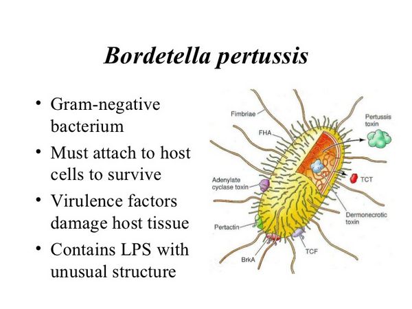
B pertussis produces a number of molecules that have been implicated in the pathogenesis of disease, including filamentous hemagglutinin (FHA), fimbriae (types 2 and 3), pertactin, pertussis toxin, adenylate cyclase toxin, dermonecrotic toxin, tracheal cytotoxin, and lipo-oligosaccharide. Among these factors, all but tracheal cytotoxin and lipo-oligosaccharide are activated by a regulatory locus referred to as the Bordetella virulence gene (Bvg) system. The roles for these factors can be considered in the context of the sequence of events involved in the pathogenesis of disease, namely entry into the host and attachment to a target tissue, persistence in the respiratory tract, and production of local damage. B pertussis does not invade beyond the respiratory mucosa. Thus systemic manifestations presumably result from dissemination of bacterial components, such as pertussis toxin.
FHA, types 2 and 3 fimbriae, pertactin, and pertussis toxin facilitate attachment to respiratory epithelium. Tracheal cytotoxin is derived from the bacterial cell wall and causes ciliostasis and sloughing of ciliated cells, thus allowing the organism to overcome mucociliary clearance. Adenylate cyclase toxin, a pore-forming cytotoxin that belongs to the RTX family, inhibits phagocyte functions, including chemotaxis, phagocytosis, oxidative burst, and bactericidal activity, and presumably enables the organism to evade local immunity. Pertussis toxin also impairs phagocyte functions, including neutrophil chemotaxis and phagocytosis. Tracheal cytotoxin, adenylate cyclase toxin, dermonecrotic toxin, and lipo-oligosaccharide may contribute to local damage to the respiratory mucosa.
Bordetella Species: Clinical Syndrome
BRANHAMELLA CATARRHALIS
Essentials of Diagnosis
- Branhamella catarrhalis is presumed to be acquired by direct contact with contaminated respiratory tract secretions or by droplet spread.
- The most common infections include otitis media, sinusitis, and, to a lesser extent, exacerbations of underlying lung disease.
- Gram stain of infected fluid shows gram-negative diplococci. Culture of infected fluids when indicated is also useful.
General Considerations
Epidemiology
B catarrhalis was previously thought to be a harmless commensal organism, but it is now clear that this organism is an important cause of human disease. B catarrhalis colonizes the nasopharynx of 5 to 10% of adults and ~ 30% of children. Colonization rates may be even higher during the winter months. B catarrhalis accounts for ~ 15% of all cases of otitis media. Evidence that B catarrhalis is a true middle ear pathogen comes from the observation that antibodies specific for this organism develop after otitis media in which a pure culture of B catarrhalis is obtained from middle ear fluid. B catarrhalis also causes sinusitis and can be recovered alone or in combination with other bacteria from direct sinus aspirates of patients with clinical and radiographic evidence of acute bacterial sinusitis.

Microbiology
The taxonomic position of B catarrhalis remains controversial. There are conflicting proposals to classify this organism as a member of the genus Moraxella, subgenus Branhamella, or to leave it as the only species in the genus Branhamella. The genera Branhamella and Moraxella are classified along with the genera Neisseria, Kingella, and Acinetobacter in the family Neisseriaceae. In contrast to Moraxella spp., which are rod-shaped, B catarrhalis appears as gram-negative diplococci with kidney-shaped cells and thus is morphologically indistinguishable from Neisseria. It grows well on blood agar and chocolate agar, forming small, opaque, gray-white colonies, 1-3 mm in diameter, which are circular and firm. Nevertheless, recovery from mucosal surfaces is facilitated by the use of selective media such as Thayer-Martin or TV broth (Mueller-Hinton broth supplemented with trimethoprim and vancomycin). Isolates typically produce cytochrome oxidase, catalase, and DNase. They are unable to ferment maltose, glucose, lactose, or sucrose, thus distinguishing them from N gonorrhoeae and N meningitidis. B catarrhalis lipopolysaccharide molecules lack long O-polysaccharide side chains and are referred to as lipo-oligosaccharide. These organisms are nonencapsulated.
Pathogenesis
The pathogenesis of localized respiratory tract disease due to B catarrhalis begins with colonization of the upper respiratory tract mucosa. Subsequently, the organism spreads contiguously to produce disease in the middle ear, the sinuses, or the lower respiratory tract. Factors that increase the likelihood of spread to the tracheobronchial tree include smoking, intercurrent viral infection, corticosteroid use, and other forms of immunosuppression. The determinants of colonization remain poorly defined but may include pili, which are present on most isolates, and a nonpilus protein called UspA1. Persistence on the respiratory mucosa may relate to the ability of the organism to resist complement-dependent bactericidal activity, which is mediated by a high-molecular-weight protein referred to as UspA2 (antigenically related to UspA1 and also called HMW-OMP). In animal models of B catarrhalis lower respiratory infection, the organism appears to release a potent chemotactic factor and elicits a striking neutrophil response in the lung.


