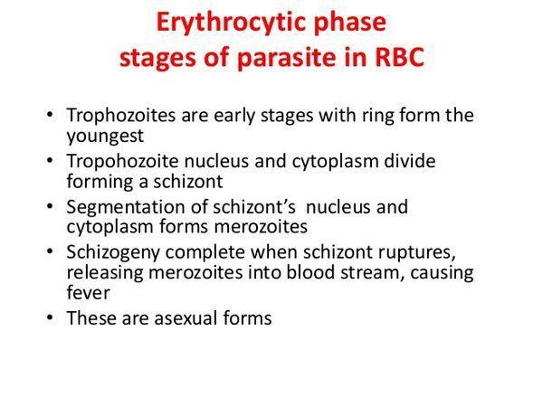
This begins with penetration into red blood cells by merozoites arising from the exoerythrocytic schizonts in the liver cells. The parasites grow rapidly, and a large central vacuole forms in the cytoplasm leading to the so-called ring form. The cytoplasm then becomes amoeboid to form the single-nucleated trophozoite. At this stage it feeds on the host cell by the process of phagotrophy during which nutrients from the red blood cell are incorporated into the parasite cytoplasm inside food vacuoles lined by a double membrane. As the hemoglobin of the RBC is metabolized the contents of the vacuoles and the RBC itself become paler, and insoluble malaria pigment (hemozoin) is formed as granules of variable size.

The trophozoite grows rapidly in size and becomes less amoeboid. New vacuoles are no longer formed. The nucleus divides by mitosis until the mature schizont is formed as a solid body containing variable numbers of nuclei. As in the exoerythrocytic schizont, discrete merozoites now form, as individual small oval or round bodies each containing a nucleus of chromatic material. The RBC ruptures, and the merozoites escape into the blood stream. Some are destroyed in the plasma, others invade red blood cells and repeat the cycle.
At some stage of the infection, merozoites entering red blood cells form sexual parasites, the male and female gametocytes, instead of repeating the erythrocytic cycle. The asexual erythrocytic cycle is completed in 2 to 3 days, depending on the species of parasite concerned, and ends with the discharge of merozoites and the destruction of the RBC. This process is called schizogony. Gametocytes take several days longer to mature. They then dwell in the individual RBC for the remainder of their existence — this may be more than 100 days. Relapse occurs only in sporozoite-induced P. vivax, P. ovale, and P. malariae infections. It does not occur in blood transfusion-transmitted infections, because there are no sporozoites. In P. falciparum infections, renewed manifestations of the malaria subsequent to the primary attack, if not resulting from fresh infection, are due to multiplication of an existing population of parasites surviving from the original infection. This is recrudescence, not relapse. A person infected with P. vivax may suffer a relapse up to 3 years after leaving an endemic area.
A nonhematologist can distinguish P. falciparum from non-falciparum malaria by these characteristics; for example; P. falciparum usually has only ring forms in the blood smear. There is a dipstick antigen-capture assay available for the diagnosis of P. falciparum malaria. The sensitivity of this test depends upon the number of parasites present. For >60 parasites per microliter it is 100% sensitive, whereas the sensitivity is 11% to 67% if there are <10 parasites per microliter. The specificity is 88 to 98%. There may only be a few parasites present on the entire blood film; so time (up to 40 to 60 minutes) and patience are needed to diagnose many cases of malaria on thin blood films. Thick blood films are useful for finding parasites but much more experience is necessary to read a thick blood film than to read a thin one.
| Table Characteristics of Various Species of Plasmodium as Observed on a Blood Film | ||||||||||||||||||||||||||||||||||||||||||||||||
|
||||||||||||||||||||||||||||||||||||||||||||||||
Clinical presentation
The initial symptoms are often referred to as “flulike” by the patient. These symptoms are non-specific and can mislead both the patient and the physician. Malaise, headache, fatigue, myalgia, and fever lead to a visit to the physician. The physical exam is usually normal. However, fever in a traveler to a malarious area should raise the suspicion of malaria. As the infection progresses the classical symptoms emerge: fever, chills, and rigors. The temperature peak may be up to ≥40°. The fever can follow a regular pattern of spikes every 2 to 3 days but it may be very irregular in P. falciparum malaria. Splenomegaly may be present. Plasmodium falciparum malaria if untreated will continue to get progressively worse. The most dreaded complication is cerebral malaria. This is manifested as a diffuse encephalopathy. The onset may be gradual or sudden, being first manifested as a generalized seizure. Coma is a poor prognostic sign and is associated with a 20% mortality rate. Adults who survive cerebral malaria usually do not have neurologic sequelae; however, about 10% of children surviving cerebral malaria, especially those with hypoglycemia, multiple seizures, and coma, have neurological deficits.
Other complications that may develop are hypoglycemia, lactic acidosis, renal failure, noncardiogenic pulmonary edema, hemolytic anemia, and thrombocytopenia. Hypoglycemia complicating malaria is seen more frequently in women and children than in men, and may be caused by failure of hepatic gluconeogenesis, increased glucose consumption by host and parasite, and drug effects (quinine and quinidine stimulate insulin secretion). Lactic acidosis commonly accompanies hypoglycemia in severe malaria, due to interference with the microcirculation by sequestered parasites leading to anaerobic glycolysis. Furthermore, the liver in these patients does not clear lactate effectively. Renal impairment is common in adults with malaria. Acute tubular necrosis requiring dialysis may occur. Noncardiogenic pulmonary edema is uncommon. The pathogenesis is unknown but it may develop even after several days of antimalarial therapy.
Prevention, control, and prophylaxis
To date no effective vaccine has been developed. Measures to reduce the frequency of mosquito bites are important. Avoid exposure to mosquitoes at peak feeding times (dawn and dusk). Use insect repellant, suitable clothing, and insecticide-impregnated bed nets. Although permethin-impregnated bed nets have reduced malarial mortality and morbidity and improved childhood growth in Africa, they may paradoxically increase the burden of disease among children, probably because infection with P. vivax very early in life protects against severe disease caused by P. falciparium malaria. Insecticides (e.g., DDT, malathione) that reduce the mosquito population have led to marked reduction in the number of cases of malaria in countries that use these insecticides.
Travelers to malarious areas are advised to take medication to prevent malaria. The choice of chemoprophylaxis depends on the area(s) to be visited, type of lodging available, duration of stay, age of the patient, the species of malaria in the area, and the drug susceptibility of these parasites. Since recommendations for chemoprophylaxis frequently change, physicians are advised to consult an up-to-date source such as the CDC Travel Information Web site (http://www.cdc.gov/travel/travel/travel12.htm).
The presence of malarial parasites in the placenta is associated with low birth weight, and maternal malarial infection during labor may be associated with a higher perinatal mortality. A review of data from randomized trials indicates that routine chemoprophylaxis for pregnant women results in a trend toward higher birth weights. Some authors suggest that chemoprophylaxis should be targeted to anemic women and primigravida.
Treatment
The drug regimens for treatment of malaria are given in Table Drugs Used to Treat Malaria in Adults and Children. If there is any doubt about whether the malarial parasites are drug-resistant, treat as if they are resistant. Even though halofantrine and mefloquine are relatively recent additions to our malaria therapeutic regimens, resistance rates to these compounds approach 50% in parts of Thailand. Primaquine is contraindicated in patients with severe G6PD (glucose-6-phosphate dehydrogenase deficiency).
A radical cure with primaquine is not indicated in areas where malaria is endemic and hence reinfection is common.
| Table Drugs Used to Treat Malaria in Adults and Children | ||||||||||||||||||||||||||||||||||||||||||||||||||||
|
||||||||||||||||||||||||||||||||||||||||||||||||||||
Chloroquine exerts its antiparasitic effects by inhibiting heme polymerase. Resistance is mediated by rapid efflux of chloroquine. Quinine and quinidine are cinchona alkaloids derived originally from the bark of the cinchona tree of South America. Quinidine is the dextrorotatory diastereomer of quinine. It is more active than quinine as an antimalarial, but it is more cardiotoxic. Both these drugs inhibit heme polymerase. Cinchonism (tinnitus, nausea, high tone deafness, and dysphoria) occurs in up to 25% of patients treated with quinine. It resolves on discontinuation of the drug. Hypotension and hypoglycemia (due to pancreatic islet cell stimulation) may occur following intravenous administration of quinine. The hypoglycemia can be reversed with octreotide. Quinine combined with tetracycline or pyrimethamine-sulfadoxine is the treatment of choice for chloroquine-resistant P. falciparum malaria. Clindamycin in combination with quinine is another treatment regimen for chloroquine-resistant P. falciparium malaria.
Mefloquine has a half-life of 20 days. It inhibits heme polymerization. The mechanism of mefloquine resistance is unknown. Children tolerate mefloquine better than adults and men tolerate it better than women. Severe neuropsychiatric reactions (psychosis, convulsions) occur in 1 in 10,000 to 1 in 13,000 when it is used in prophylactic doses; with treatment doses (15 mg/kg) such reactions occur 1 in 215 to 1 in 1700 persons treated. This drug is contraindicated in those who have a history of seizures or major psychiatric disorder.
Halofantrine is a phenanthrene methanol derivative related to mefloquine and quinine. The mechanisms of action and resistance are unknown. Retreatment on day 7 is necessary to prevent recrudescence of infection. High doses of halofantrine (8 mg/kg every 8 hours for 3 days) have low failure rates, but cardiotoxicity, sometimes fatal, occurs — the progesterone receptor and QT intervals are prolonged. Halofantrine should never be used in combination with mefloquine or quinine because these combinations are additive in terms of QT prolongation. Halofantrine should not be ingested with fatty foods. It should be used only when other treatment options are contraindicated or inappropriate, and the dose should be limited to 8 mg every 6 hours times 3 doses, and repeated in 1 week.
Artemisinin (ginghaosu) was isolated in 1972 from Artemisia annua, a plant used by traditional Chinese practitioners to treat fever. It is available as the parent compound artemisinin, and as three semisynthetic derivates, artesunate for oral administration, artemether, and arteether for intramuscular injection. Qinghaosu and its derivatives result in faster parasite clearance than other antimalarials. They are as effective as quinine in the treatment of severe and complicated malaria. There is in vitro synergy with mefloquine and tetracycline.
Atovaquone (a hydroxynaphthoquinone used as an alternative agent to treat Pneumocystis carinii pneumonia) is effective against multidrug–resistant P. falciparum malaria. It cannot be used alone, but, in combination with doxycycline or proguanil, cure rates of >90% against MDR P. falciparum are obtained. Malarone is a combination of atovaquone (250 mg) and proquanil (100 mg). Four tablets once daily for three days has been effective in the treatment of MDR P. falciparum.
P. vivax malaria acquired in Papua, New Guinea; Thailand; or Oceania may relapse after standard courses of primaquine. These patients should be retreated with twice (30 mg/kg) the standard dose for 14 days.
Severe P. falciparum malaria (often cerebral malaria) can be treated with a continuous infusion of quinidine and exchange transfusion. Exchange transfusion should be considered when any one of the following are present in severe P. falciparum malaria: >10% of red cells parasitized; disseminated intravascular coagulation; acute renal failure.
Corticosteroid therapy is of no benefit and indeed it is deleterious in the treatment of cerebral malaria. Acetaminophen (in children) has no antipyretic benefits over mechanical antipyresis and it prolongs the time of parasitic clearance in P. falciparum malaria by 16 hours.


