Essentials of Diagnosis
- Stool examination reveals spheroidal yellow-brown eggs (31-43 mm).
- Motile proglottids that appear singly in stool.
- Mature proglottids are square.
- Scolex has no hooklets and four suckers.
- Gravid proglottid has 15-20 lateral branches.
General Considerations
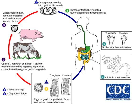
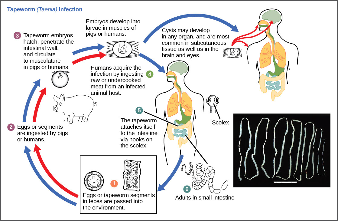
T saginata infection is commonly associated with the ingestion of undercooked beef. This is distinguished from infection with T solium because human infection with the larval form (as in cysticercosis) is extremely rare with T saginata infection. T saginata infection is common in areas of the world with intensive cattle breeding, such as central Asia and central and eastern Africa. Alternative intermediate hosts for T saginata include llamas, buffalo, and giraffes. The life cycle for T saginata is similar to that of T solium; larvae are ingested in infected meat, and the tapeworm attaches to the intestinal epithelium and matures in 12 weeks. Mature tapeworms produce gravid proglottids with characteristic 15-20 lateral branches, which contain numerous eggs. Ingestion of eggs or proglottids by cows leads to hatching of eggs, and larvae that migrate into striated muscle. Case reports exist about T saginata cysticercosis in humans, although the incidence is exceedingly uncommon.
Clinical Findings
Signs and Symptoms
Infection with T saginata is most often asymptomatic, although a minority of patients may report nonspecific abdominal cramps or malaise. The proglottids of T saginata are motile, and patients may report seeing moving segments in the stool.
Laboratory Findings
Examination of the blood in patients with T saginata infection typically reveals no abnormalities, although a mild leukocytosis with eosinophilia may be present. Otherwise all laboratory tests except the microscopic stool examination will be normal. The stool examination will frequently reveal eggs and proglottids. The main basis for differentiating T saginata from T solium is the gravid proglottid, which for T solium has 7-13 lateral branches on each side of the uterus, whereas T saginata has 15-20 lateral branches.
Differential Diagnosis
Infection with T saginata is usually not associated with clinical symptoms. Patients most often seek medical attention after finding T saginata proglottids in stools or on clothing. The main differential diagnosis is to differentiate T saginata proglottids from T solium proglottids. If no gravid proglottids are present, then differentiation may not be possible, in which case patients should be treated as though they have infection with T solium.
Complications
Usually no complications are associated with T saginata; however, regurgitation and aspiration of proglottids may occur.
Treatment
Therapy for infection with T saginata is similar to treatment of intestinal T solium, a single dose of either praziquantel or niclosamide (see Box 2). Follow-up examinations of stool should be performed 1 month after treatment.
Prognosis
The prognosis for patients with intestinal T saginata infection is excellent.
Prevention & Control
Prevention of infection with T saginata involves thorough cooking of beef and beef products to > 65 °C core temperature (Box 3). Beef should also be inspected for the presence of cysts, and infected carcasses destroyed.


