Essentials of Diagnosis
- Epidemic systemic viral illness, primarily of children and young adults.
- Exanthematous disease with fever, cough, coryza, and conjunctivitis.
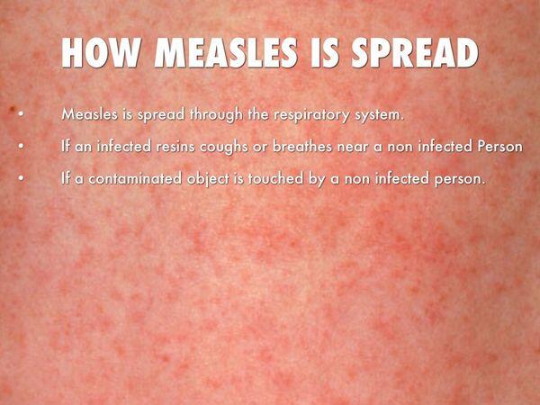
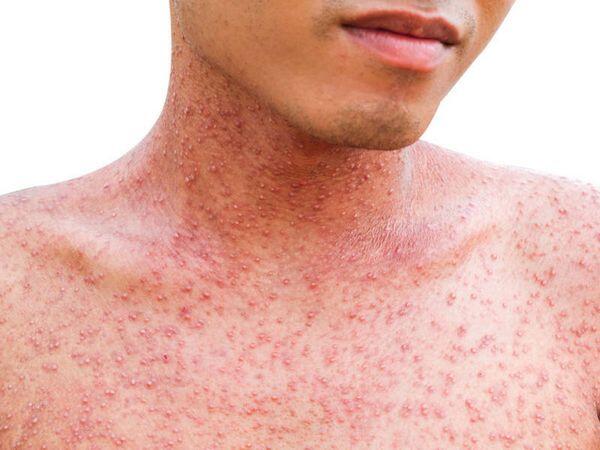
- Exanthem is a maculopapular, confluent rash that is centrifugally spread from the head to the extremities.
- Koplik’s spots are a pathognomonic enanthem that occurs on the buccal mucosa.
- Incidence has drastically dropped in the postvaccination era.
General Considerations
Epidemiology
Rubeola, commonly known as measles, is a virus spread primarily in the winter and early spring. Like mumps and rubella, vaccination has drastically changed the epidemiology of measles. In the prevaccination era, rubeola followed a biannual epidemic cycle. It is prevalent throughout the world.
Measles vaccine was introduced for commercial use in 1963. From 1963 to 1967, ~900,000 children received the inactivated measles vaccine. The attenuated, live-virus vaccine was introduced in the late 1960s. By 1973, the inactivated vaccine was no longer available. Before 1963, between 200,000 and 600,000 cases were reported annually in the United States. However, even these reports represent only a fraction of the actual case rate. The number of reported cases dropped from 22,231 in 1968 to 1,497 in 1983.
In the mid-1980s a resurgence of reported cases occurred primarily in the preschool population. Nearly 19,000 cases were reported in 1989. The majority of these cases were in vaccine-eligible, unvaccinated preschool children living in urban areas. In the late 1980s and early 1990s several outbreaks occurred among adolescents and young adults, primarily on college campuses. Most of the cases were in patients with a history of receiving only a single dose of the vaccine. Since 1993, < 1,000 cases have been reported per year. This is likely owing to aggressive revaccination efforts. A second measles vaccination is now standard at ages 4-6 years.
Microbiology
Measles is caused by a pleomorphic 100- to 250-nm RNA virus of the Paramyxoviridae family. It is a member of the genus Morbillivirus. The virus has three structural proteins complexed within its helical nucleocapsid. The lipoprotein envelope contains three proteins (F, H, and M). The F glycoprotein facilitates host cell adhesion and penetration. The H glycoprotein is a hemagglutinin. The M protein is a nonglycosylated protein that lines the inner lipid bilayer and facilitates viral maturation. The virus is labile and sensitive to heat, ultraviolet (UV) light, and extremes of pH.
Pathogenesis
Measles virus is spread primarily via respiratory droplet nuclei. Direct contact is also a mode of transmission. Humans are the only natural reservoir for the virus. The primary site of adhesion and invasion is the respiratory epithelium, with quick spread to lymphatic tissues and subsequent viremia.
Measles virus is communicable for 3-5 days before the outbreak of the rash. It remains communicable up to 4 days after the appearance of rash. Immunocompromised patients can shed virus for an extended period of time. Patients who develop subacute sclerosing panencephalitis do not continue to shed virus.
CLINICAL SYNDROMES
TYPICAL MEASLES
Clinical Findings
Signs and Symptoms
Typical measles, also referred to as “natural measles,” can occur at any age (Table 1). Immunity is lifelong, so recurrence is extremely rare. A prodromal phase of 2-4 days is marked by fever, malaise, cough, coryza, conjunctivitis, and pharyngitis. A pathognomonic enanthem of measles (Koplik’s spots) erupts on the buccal mucosa during this phase. These appear as small blue to white plaques.

The exanthem phase begins ~2 weeks after exposure. The maculopapular rash first appears on the brow and posterior auricular area. It progresses to cover the face and spreads to the distal extremities within 72 h. Large areas of the rash will progress to confluence, then desquamate leaving transient hyperpigmented areas. The rash usually begins to clear on the 3rd or 4th day. The fever peaks on the 2nd or 3rd day of the rash.
Laboratory Findings
The virus can be isolated from a nasopharyngeal swab, conjunctival swab, blood, and urine during the febrile course of the illness. Isolation of the virus can be technically difficult. A variety of tissue cultures can be used to grow the virus. Human and monkey kidney cell lines are typically used with clinical specimens. Acute and convalescent serologies can be helpful. Serologies should be drawn 2-4 weeks apart to monitor progression. Immunoglobulin M (IgM) is detectable from 3 to 30 days after the onset of rash. Measles antibody is detectable in the cerebrospinal fluid of patients with subacute sclerosing panencephalitis. These patients often have a high IgG titer in the serum as well.
Imaging
Chest radiographs may demonstrate diffuse infiltrates if pneumonia is present.
Differential Diagnosis
The differential diagnosis of measles includes Kawasaki disease, Stevens-Johnson syndrome, and other viral exanthems.
Complications
Acute complications can include meningoencephalitis, pneumonia, otitis media, and laryngotracheitis (Box 1). Measles meningoencephalitis occurs in 1 per 1000 cases. It has a high morbidity and mortality rate. “Black measles” is a severe, hemorrhagic variation of typical measles. It is extremely rare in the postvaccination era. It has a high mortality rate. Patients present with a confluent hemorrhagic skin rash, encephalitis, and pneumonia. Bleeding often occurs via nose, mouth, and gastrointestinal tract.
Measles infection during pregnancy produces significant fetal morbidity and mortality, especially if infection occurs during the first trimester. Measles uncommonly produces severe abdominal pain during the acute febrile phase. Etiologies for this pain can be mesenteric lymphadenitis or appendicitis. Evidence of peritonitis warrants prompt surgical evaluation.
Subacute sclerosing panencephalitis (SSPE) is a rare, late-onset, lethal neurodegenerative sequela of measles infection. It occurs with an incidence of 0.6-2.2 cases/100,000 infections and 1 case/1 million vaccinations. SSPE results from a slowly progressive chronic infection with the virus. SSPE becomes clinically evident an average of 7 years after initial measles exposure. Symptoms include unusual behavior, developmental regression, ataxia, myoclonic jerks, visual impairment, and aphasia. All of these symptoms are progressive, leading ultimately to decorticate rigidity and death, which usually occurs 6-9 months after onset of symptoms. Confirmation of the diagnosis can be made by electroencephalogram, serology, and analysis of the cerebrospinal fluid. Cerebrospinal fluid shows high IgG as does serum. No current therapy is effective.

MODIFIED MEASLES
Modified measles occurs in individuals receiving an incomplete live virus vaccination series and subsequently being infected by the natural virus. It can also occur in infants under 9 months owing to the presence of maternally derived antibodies. It is similar to natural measles infection but less severe (see Table 1). The prodromal phase may be shorter and less severe. Koplik’s spots may be scant or absent. The exanthem rash usually does not progress to confluence, but has a similar duration. Viral isolation is unaffected. All the complications and sequelae of typical measles can occur.
ATYPICAL MEASLES
Atypical measles occurs in some individuals who received the inactivated virus vaccine in the 1960s and were subsequently exposed to the natural virus. These patients have a prodromal phase noted by sudden onset of high fever, headache, myalgia, and abdominal pain (see Table 1). Lobar pneumonia with effusion is common. These patients rarely have Koplik’s spots. The exanthem phase begins on the 2nd or 3rd day of illness. The maculopapular rash begins on the palms and soles and spreads centrally. It can progress to vesiculation. Urticaria, purpura, and petechiae are common.
Chest radiographs will commonly show lobar consolidation. Acute and convalescent titers and viral culture are useful. The differential diagnosis of this exanthem includes rickettsial disease, meningococcemia, and hemorrhagic fever.
Diagnosis
Diagnosis of measles is usually based on physical findings. Finding Koplik’s spots and monitoring the usual progression of the exanthem are helpful clues. An attempt should be made to culture the virus during the febrile course of the illness. Culture can often be technically difficult. Serology should be obtained as well. Confirmation of a diagnosis should be promptly reported to a local government health agency for epidemiologic tracking.
Treatment
Treatment of measles is often supportive. Hospitalization is not warranted unless the patient is dehydrated, in respiratory distress, encephalopathic, or otherwise compromised. Young patients requiring hospitalization (Box 2) should receive vitamin A. Recent clinical studies have shown a decrease in morbidity and mortality in patients receiving vitamin A supplementation. Patients of any age may benefit from vitamin A supplementation if their cases are clinically severe. All patients in areas of known vitamin A deficiency (ie, Third World countries) should receive supplementation if measles is endemic to the area.
Immune globulin may be used to protect susceptible contacts of confirmed measles cases. Immune globulin must be used within 6 days of exposure for maximum efficacy. Contacts who received at least one dose of measles vaccine at age = 12 months do not need immune globulin if they are immunocompetent. Candidates for immune globulin would include nonimmunized pregnant women, the immunocompromised, and infants < 1 year of age. Infants < 5 months of age do have passive maternal antibodies if their mother can be confirmed to be measles immune. If maternal immunity is confirmed, these infants do not need immune globulin. If children receive immune globulin, they should not receive measles vaccine for 5 months (if receiving 0.25 cc/kg immune globulin) or 6 months (if receiving 0.5 cc/kg immune globulin).
The live virus vaccine can modify the infection in a nonimmunized person, if given within 72 h of exposure. Clinical response varies. Usual vaccine contraindications apply to the vaccine if used in this modality (see Box 2).
Prevention & Control
Measles is highly communicable. Any hospitalized patient suspected of harboring the virus should be in droplet isolation. Good hand washing and appropriate handling of fomites should be observed as well. This isolation should be maintained during the prodromal phase and 4 days from the onset of the exanthem. Children should also be excluded from school and daycare during this period. Isolation may need to be extended in complicated cases. Some immunocompromised patients can shed virus for an extended period.
School quarantine issues are often difficult. Virus is shed and communicable in the prodromal phase. The diagnosis of measles is often not suspect in this phase. The index case may have many contacts before diagnosis. Quarantine issues should be addressed individually and in conjunction with a local government health agency.
Live attenuated measles vaccine should be administered to healthy individuals at two intervals. The first dose is recommended at age 12-15 months. The second dose should be at age 4-6 years. Children who miss the second dose should receive it before age 12 years.
In epidemic conditions, a dose may be given from ages 6 to 11 months. These patients will still need a dose at age 12-15 months and at 4-6 years (Box 3).
Table 1. Measles clinical findings.
Typical Measles
Modified Measles
Atypical Measles
Cause
- Natural measles infection
- Rare
- Incomplete live-virus vaccination series followed by natural measles infection
- Rare
- Inactivated measles vaccination, followed by exposure to natural measles virus
Incubation
- 8-12 d
- Similar to typical measles
- 1-2 weeks
Prodromal phase
- 2-4 days duration
- Symptoms progressive
- Fever & malaise
- Cough & coryza
- Conjunctivitis
- Pharyngitis
- Koplik’s spots (pathognomonic enanthem of measles) appear on days 9-11; blue to white spots on the lower buccal mucosa
- Similar prodromal symptoms, but less severe and shortened duration
- Koplik’s spots few to absent
- Sudden onset of high fever
- Headache & myalgia
- Abdominal pain
- Koplik’s spots rare
Exanthem phase
- Rash begins 2 weeks
- Fever peaks on day 2 or 3 after exposure of rash
- Erythematous maculopapular rash which progresses to confluence
- Initially involves the forehead & posterior auricular area
- Rash spreads from head to feet in 48 to 72 h
- Rash begins to clear on day 3 to 4
- Clears from head to feet
- Confluent areas desquamate, leaving brown, hyperpigmented areas
- Similar rash morphology and progression
- No confluence
- Rash appears on second or third day of illness
- Initially erythematous, maculopapular, and spreading from distal extremities to the head
- Present on palms and soles
- Can progress to vesiculation
- Urticaria, purpura, & petechiae are common
- Lobar pneumonia with effusion is common
BOX 1. Complications of Measles
More Common
- Meningoencephalitis
- Pneumonia
- Otitis media
- Laryngotracheitis
Less Common
- Subacute sclerosing panencephalitis
- Myocarditis/pericarditis
- “Black measles”
- Thrombocytopenia purpura
- Stevens-Johnson syndrome
- Mesenteric lymphadenitis
- Appendicitis
BOX 2. Treatment of Measles
Indication
Dosage
Supportive Care
- Vast majority of cases
- Antipyretics/analgesics
- Fluid
Vitamin A
- Patients 6 months to 2 years of age requiring hospitalization
- Immunodeficiency
- Ophthalmologic evidence of vitamin A deficiency
- Impaired intestinal absorption
- Malnutrition
- Immigration from an area of high measles mortality
- Children >1 year: 200,000 IU orally
- Children 6 months to 1 year: 100,000 IU orally
- Repeat dose at 24 h and 4 weeks if clinical evidence of vitamin A deficiency
Immune Globulin
- Susceptible contacts of confirmed cases, especially infants, pregnant women, and immunocompromised
- Infants under 5 months have passive immunity if mother has confirmed immunity
- Must be used within 6 days of exposure
- 0.25 cc/kg IM
- 0.5 cc/kg IM for immunocompromised
- Max dose = 15 cc
Live-Virus Vaccine
- Indicated for susceptible contacts who are greater than 6 months of age, not pregnant, and immunocompetent
- Must be used within 72 h of exposure
- 0.5 cc SQ
- Clinical response variable when used postexposure
Ribavarin
- Severe cases
- Immunocompromised
- Not FDA approved for measles treatment
- No controlled clinical trials/TR>
BOX 3. Control of Measles
Vaccine
- Live attenuated measles vaccine is administered 0.5 cc SQ. Commonly given in combination with MMR (measles, mumps, rubella) vaccine
- First dose is recommended at from 12 to 15 months of age
- Second dose is recommended at school entry (age 4 to 6 years)
- If no preschool dose is given, the second dose should be administered before age 12
- Improperly stored vaccine may result in failure
- A dose may be given to infants age 6 to 11 months during epidemic conditions; these infants must still follow the two-dose guideline above
MMR Vaccine Contraindications
- Pregnancy
- Febrile illness
- Planned pregnancy within 3 months
- Severe immunocompromised state
- Blood product or immune globulin within 3 to 6 months (dose dependent)
- Anaphylaxis to neomycin
MMR Vaccine Should Be Used with Caution in These Situations
- Seizure disorder
- Thrombocytopenia
- Egg allergy
Isolation Precautions
- Respiratory isolation required during the prodromal phase through 4 days from the onset of rash in healthy patients
- Immunocompromised patients usually shed virus for an extended period

