Essentials of Diagnosis
- Multiple, large, eosinophilic cytoplasmic inclusions in epithelial cells (H & E stain).
- Vesicular or nodular skin lesions.
- Exposure by occupation or close personal contact.
- High index of suspicion in patients with skin lesions and exposure to sheep, goats, or cows.

General Considerations
Poxviruses are a large, complex family of viruses that cause disease in humans and other animals (Table 1). Of the many genera in this family, only species of Orthopoxvirus and Molluscipoxvirus are associated specifically with humans. The former contains variola virus (smallpox), which is currently of historical interest only, and the latter molluscum contagiosum virus. Poxviruses in other genera naturally infect animals (zoonoses) but cause incidental infection of humans.
Epidemiology
Molluscum contagiosum infection is spread through direct person-to-person contact or through sharing of common towels etc. The agents of the zoonotic poxvirus diseases spread by direct contact.
Microbiology
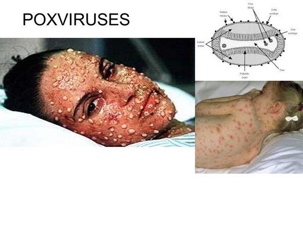
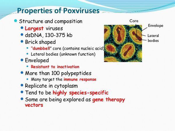
Poxviruses are the largest of all viruses, measuring 230 by 300 nm, and are ovoid to brick shaped. They have a capsid that is referred to as complex because it has neither helical nor icosahedral symmetry. An outer membrane and envelope enclose the core and core membrane. The viral genome is a single strand of large, linear, double-stranded DNA (molecular weight approximately 100 × 106-100 × 2006).
Replication of poxviruses is unique among DNA-containing viruses, because the entire multiplication cycle takes place within the host cell cytoplasm. Viral penetration occurs within phagocytic vacuoles. Uncoating of the outer membrane occurs in the vacuole. Early gene transcription occurs within the viral core. Among the early proteins produced is an uncoating protein that removes the core membrane, liberating viral DNA into the cell cytoplasm. Replication of viral DNA follows in electron-dense cytoplasmic inclusions, referred to as factories. Late viral mRNA is translated into structural proteins, which are glycosylated, phosphorylated, and cleaved before assembly. Unlike other viruses, poxvirus membranes form de novo in the cytoplasm, rather than as part of a host cell membrane that is picked up during a budding process. About 10,000 viral particles are produced per infected cell.
C. Pathogenesis. The pathologic hallmark of poxviruses is cell proliferation, manifested by the skin lesions that give this family of viruses its name. Most of the viruses of current human importance are primary pathogens in vertebrates other than humans (eg, cow, sheep, and goats) and infect humans only through accidental occupational exposure (zoonosis). As stated earlier, the exception to this is molluscum contagiosum.
CLINICAL SYNDROMES
MOLLUSCUM CONTAGIOSUM
Clinical Findings
Signs and Symptoms
The incubation period is 2-8 weeks. The lesions differ significantly from pox lesions in that they are nodular to wartlike. They begin as papules and progress to pearly, umbilicated nodules 2-10 mm in diameter, with a central caseous plug that can be readily expressed. They are most common on the trunk, genitalia, and proximal extremities and usually occur in a cluster of 5-20 nodules (Box 1). Lesions disappear in 2-12 months even without treatment. Often a history can be elicited of contact with an individual with skin lesions, eg, from wrestling or sexual contact.
Laboratory Findings
The lesions of molluscum contagiosum are caused by a poxvirus that is unclassified because it does not grow in cell cultures but can be confirmed histologically by finding large eosinophilic inclusions in the cytoplasm (molluscum bodies) of epithelial cells.
Differential Diagnosis
Molluscum contagiosum lesions may be mistaken for warts but can be distinguished by expression of cheesy material from the center of each lesion, by histologic examination, and by location.
ZOONOTIC POXVIRUSES
ORF & COWPOX
The poxviruses of animals such as sheep or goats (orf) and cows (cowpox) can infect humans, usually as a result of accidental direct contact.
Clinical Findings
Signs and Symptoms
Single or multiple nodular lesions are usually found on the fingers or face and are vesicular or pustular (cowpox) or granulomatous (orf). After progressing from a vesicular lesion to a nodular mass, the lesion usually regresses in 25-35 days.
Laboratory Findings
There are no characteristic abnormalities in blood counts or chemistries.
Differential Diagnosis

The lesion may be mistaken for anthrax. The anthrax lesion, however, is vesicular, then ulcerative, and finally, a black eschar and does not develop a nodular appearance.
Complications
Because orf and cowpox lesions resolve completely, there are no complications.

PSEUDOCOWPOX OR MILKER’S NODULES
Clinical Findings
Signs and Symptoms
Pseudocowpox or Milker’s nodules is a cutaneous disease of cattle, distinct from cowpox, that can cause localized papular lesions which progress to purplish nodules. Healing of the skin lesions may take 4-8 weeks.
Differential Diagnosis
The lesions can resemble the eschar of anthrax or the early lesion of orf or cowpox, but milker’s nodules are nonulcerating.
MONKEYPOX
Over 100 cases of an illness resembling smallpox have been attributed to the monkeypox virus. Multiple vesiculopustular lesions occur, but the illness is not as severe. All have occurred in western and central Africa, especially Zaire. There has been concern that this agent might replace the smallpox virus and become epidemic in humans, but this has not materialized probably because animal poxviruses seem highly adapted to their particular host and require very close, usually direct, contact for transmission.
Treatment
Molluscum Contagiosum. Treatment of molluscum contagiosum, if lesions are extensive or cosmetically disfiguring, is curettage, forceps removal of central core, or application of liquid nitrogen or iodine solutions (Box 2).
Zoonotic Poxviruses. Since the lesions of zoonotic poxviruses heal in 4-6 weeks, treatment is unnecessary.
Prevention & Control
Molluscum Contagiosum. Prevention of molluscum contagiosum consists of avoiding direct or indirect contact with individuals exhibiting characteristic skin lesions (Box 3).
Zoonotic Poxviruses. The zoonotic poxviruses can be avoided by avoiding contact with animals exhibiting skin lesions, especially when the human has potentially infected cuts or abrasions.
Table 1. Diseases associated with poxvirus.
Virus
Disease
Source
Location
Variola
Smallpox (now extinct)
Humans
Extinct
Vaccinia
Vaccination complications
Vaccine
Research laboratories
Molluscum contagiosum
Many skin lesions
Humans
Worldwide
Orf
Localized lesion
Zoonosis: sheep, goats
Worldwide
Cowpox
Localized lesion
Zoonosis: cows
Europe
Pseudocowpox
Milker’s nodule
Zoonosis: dairy cows
Worldwide
Monkeypox
Generalized disease
Zoonosis: monkeys, squirrels
Africa
BOX 1. Molluscum Contagiosum Infection
Children
Adults
More Common
Lesions on exposed epithelial surfaces
Genital lesions, may be generalized in AIDS patients
Less Common
BOX 2. Treatment of Molluscum Contagiosum Infection
Children
Adults
First Choice
No systemic treatment
No systemic treatment
Second Choice
- Local Rx with liquid nitrogen or iodine solution
- Laser, cryotherapy, curettage, especially in AIDS patients
Pediatric Considerations
Rx rarely necessary
BOX 3. Control of Molluscum Contagiosum Infection
Prophylactic Measures
In adults, may be sexually transmitted and “safe sex” may decrease spread.
Direct contact with lesions should be avoided.
Isolation Precautions
None required


